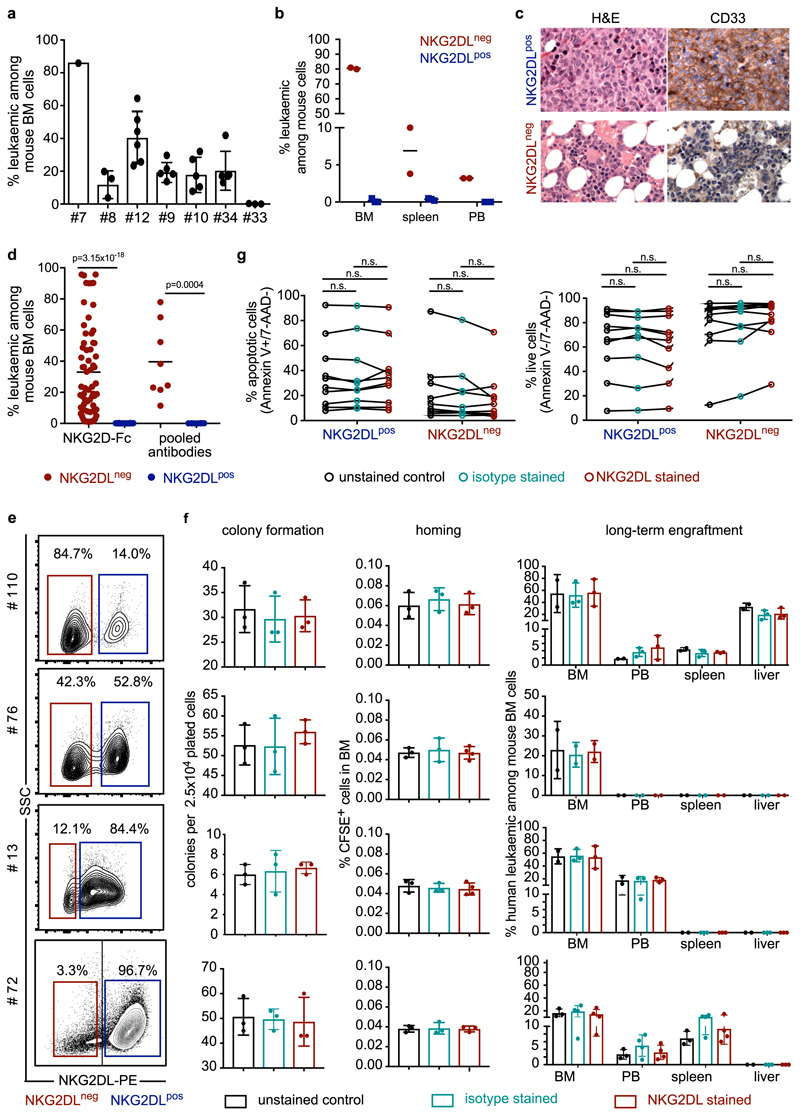
Extended Data Fig. 2. Only NKG2DL− subpopulations of AML cells are serially re-transplantable and selectively engraft NSG mice after intrafemoral injection.
a–c, Bone marrow cells isolated from mice transplanted with NKG2DL− AML cells were re-transplanted into sublethally irradiated secondary recipient mice, either unsorted (a, n = 7 cases of AML) or after sorting into NKG2DL− and NKG2DL+ AML cells (b, c, no. 1) (see Supplementary Table 2). Flow cytometric analyses in secondary recipients are shown, indicating percentages of engrafted human AML cells among total bone marrow cells at 12 (a) or 15 (b) weeks after transplantation. Each dot represents one transplanted mouse; n = 2 mice for NKG2DL−; n = 3 mice for NKG2DL+. c, Histological analyses of mice (H&E and anti-CD33 show exemplary data, no. 1; two analysed mice for NKG2DL− and three for NKG2DL+ cells). The NKG2DL staining procedures do not affect the results. d, NKG2DL− and corresponding NKG2DL+ cells were sorted either using NKG2D–Fc (n = 65 mice for NKG2DL−; n = 79 mice for NKG2DL+; n = 12 cases of AML) or using pooled antibodies against individual NKG2DLs (MICA, MICB, ULBP1 and ULBP2, ULBP5 or ULBP6; n = 3 cases of AML; n = 8 mice per group) and then transplanted at equal numbers for each case of AML intrafemorally in pre-irradiated NSG mice. For detailed mouse numbers per patient and subpopulation, see Supp lementary Table 2. Human leukaemic engraftment in mouse bone marrow was assessed 12–16 weeks after transplantation; percentages are shown of human leukaemic among mouse bone marrow cells in mice transplanted with NKG2DL− or NKG2DL+ subpopulations for each case of AML. Centre values represent means; each dot represents one mouse; a Student’s t-test was used for statistical analysis. Note that these samples are also included in the summarized analysis shown in Fig. 1g. e, AML cells with variable expression of NKG2DLs were stained with NKG2D–Fc, isotype or empty control and analysed side-by-side without prior sorting. f, Quantifications of results obtained in colony formation (left), homing (middle) (n = 3 mice per group and patient sample, except for n = 4 mice in NKG2D–Fc-stained group with patients no. 13 and 76) and in vivo long-term engraftment assays in NSG mice (right). Mice per patient sample and condition: no. 110, 3 NKG2D–Fc, 3 isotype and 2 empty control; no. 76, 2 NKG2D–Fc, 2 isotype and 2 empty control; no. 13, 3 NKG2D–Fc, 3 isotype and 2 empty control; and no. 72, 4 NKG2D–Fc, 4 isotype and 3 empty control. g, Annexin V and 7-AAD stainings indicating apoptotic (left) and live (right) cells among AML cells incubated for 24 h with NKG2D–Fc, isotype or control medium (n = 10 cases of AML). f, Two-sided Kruskal–Wallis test was used for statistical analysis. Centre values represent mean, error bars represent s.d.

