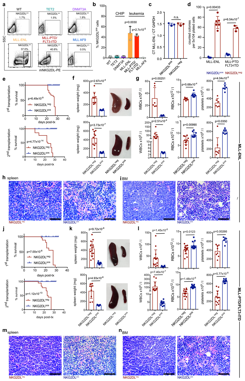
Extended Data Fig. 4. Expression of NKG2DLs on mouse leukaemic cells and association with a lack of leukaemogenicity in syngeneic mouse models.
a, b, Staining of whole bone marrow cells derived from healthy wild-type (WT), pre-leukaemic (chromatin immunoprecipitation, Dnmt3a or Tet2 mutated) or leukaemic (Mll-Enl, Mll-ptd/Flt3-ITD and Mll-Af9) mice using a mouse NKG2D tetramer. Representative flow cytometric plots (a) and summarized quantifications of NKG2DL+ cells among total mouse bone marrow cells (b) (n = 3 mice per model and condition; a two-sided Student’s t-test was used for statistical analysis). c–m, NKG2DL− and corresponding NKG2DL+ cells were sorted from leukaemic Mll-Enl and Mll-ptd/Flt3-ITD mice and analysed by qRT–PCR for the genetic leukaemic driver (c) (Mll-Enl mice, n = 3), and at equal numbers in colony-forming assays (d) (n = 6 for Mll-Enl mice; n = 3 for Mll-ptd/Flt3-ITD mice). e–n, In vivo primary and secondary transplantation (tx) assays. e, j, Kaplan–Meier-survival analyses (n = 24 mice for primary transplantations; n = 18 mice for secondary transplantations; log-rank Mantel–Cox test was used for statistical analysis). f, k, Spleen assessment with representative images and weight quantification (f, Mll-Enlmice, primary transplantation, n = 9 NKG2DL− and n = 12 NKG2DL+; secondary transplantation, n = 10 NKG2DL− and n = 12 NKG2DL+; k, Mll-ptd/Flt3-ITD mice, primary transplantation, n = 15 NKG2DL− and n = 13 NKG2DL+; secondary transplantation, n = 9 NKG2DL− and n = 11 NKG2DL+). g, l, Peripheral blood counts (g, Mll-Enl mice, primary transplantation, n = 6 NKG2DL− and n = 6 NKG2DL+; secondary transplantation, n = 10 NKG2DL− and n = 9 NKG2DL+; l, Mll-ptd/Flt3-ITDmice, primary transplantation, n = 14 NKG2DL− and n = 12 NKG2DL+; secondary transplantation, n = 11 NKG2DL− and n = 11 NKG2DL+). RBCs, red blood cells; WBCs, white blood cells. h, m, i, n, Representative histopathological analyses: spleen (h, m), bone marrow (i, n). H&E. Scale bars, 50 μm. A two-sided Mann–Whitney U test (g, l, WBCs, primary transplantation; g, platelets, secondary transplantation) or a two-sided Student’s t-test (all other analyses) were used for statistical analysis. Centre values represent mean, error bars represent s.d. for all plots.

