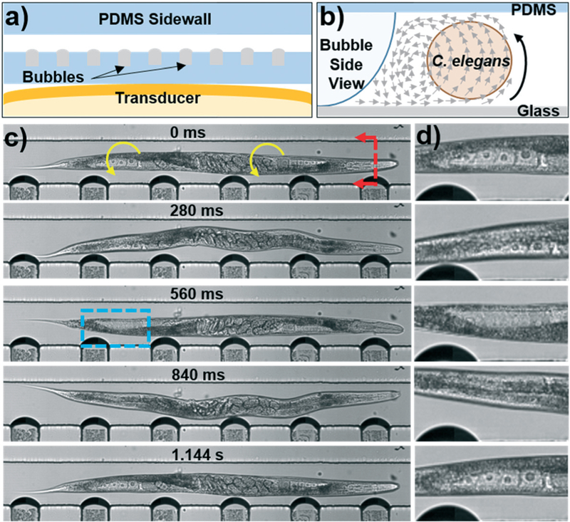
Fig. 3.
a) Schematic of the bubble-based acoustofluidic device. b) Schematic of out-of-plane streaming used to rotate C. elegans. c) Photos taken throughout the rotation of a C. elegans. A 20 V, 19.1 kHz signal was applied to the transducer to induce bubble vibration and C. elegans rotation. The blue box indicates the location of the (d) close-up of the proximal distal gonad provided on the right hand side of each photo. Oocytes can be seen clearly as they rotate into and out of the focal plane of the camera.

