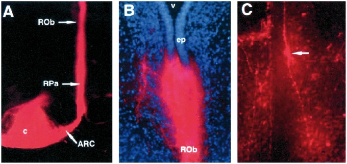
FIGURE 4.
Labeling of the caudal raphe from the arcuate nucleus. (A) Placement of DiI crystal in the arcuate nucleus (horizontal plane) with labeling of the caudal raphe, including the raphe pallidus (RPa) and the raphe obscurus (ROb). (B) Single DiI-labeled dorsoventral fiber in the ROb from the arcuate nucleus (×76). (C) Di-labeled fusiform cell body (arrow) (dorsal process and ventral process in the ROb) ×76. Reprinted from (69) with permission from Oxford University Press.

