FIGURE 2.
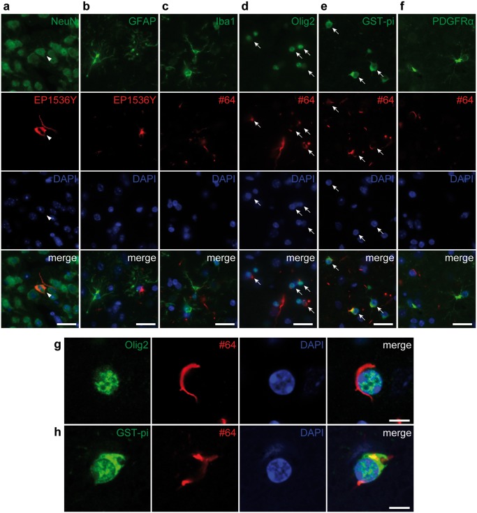
Alpha-synuclein pathology is observed mostly in neurons and mature oligodendrocytes in the ipsilateral piriform cortex at 18 months after injections of mouse α-Syn preformed fibrils into the olfactory bulb. Double label immunofluorescence images. (A) Neuronal nuclear antigen (NeuN) (green) and phosphorylated α-Syn (pSyn) (EP1536Y, red). pSyn-positive pathology in a NeuN-positive neuron (arrowheads). (B) Glial fibrillary acidic protein (green) and pSyn (EP1536Y, red). (C) Ionized calcium-binding adapter molecule 1 (green) and pSyn (#64, red). (D) Oligodendrocyte transcription factor 2 (Olig2) (green) and pSyn (#64, red). pSyn-positive pathology is seen in Olig2-positive oligodendrocytes (arrows). (E) Glutathione S-transferase pi (GST-pi) (green) and pSyn (#64, red). pSyn-positive pathology in GST-pi-positive mature oligodendrocytes (arrows). (F) Platelet-derived growth factor receptor α (green) and pSyn (#64, red). (G) Confocal microscope images of Olig2 (green) and pSyn (#64, red). (H) Confocal microscope images of GST-pi (green) and pSyn (#64, red). Scale bars: 20 µm (A–F), 5 µm (G, H).

