Figure 3.
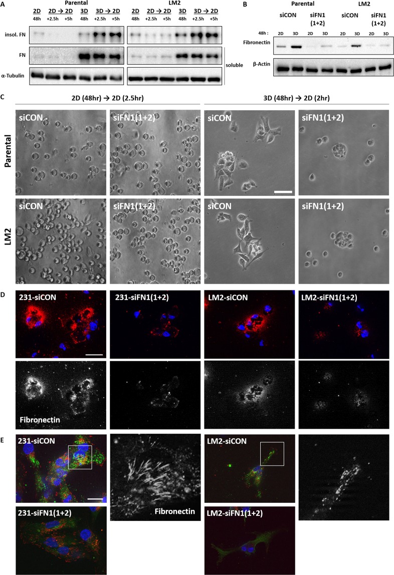
Increased fibronectin of MDA-MB-231 parental and LM2 cells in 3D facilitates cell attachment on 2D. Cells grown in 2D or 3D suspension cultures for 48 hours were re-cultivated in serum-free culture media on 2D culture plates and subjected to immunoblot after being harvested at 2.5 hr and 5 hr from 2D re-cultures and DOC-insoluble and DOC-soluble fibronectin was detected (A). DOC-soluble fibronectin used as internal loading control for comparison. DOC-soluble and DOC-insoluble lysates were loaded into different gels and cropped for immunoblots. (B) Protein expression changes were observed after FN1 siRNAs were treated in MDA-MA-231 parental and LM2 cells. The mixture of two different kinds of siRNAs against FN1 were transfected into cells in a serial manner in which 25 nM siRNAs were treated each time, and totally 50 nM siRNAs were treated. The blots for fibronectin and α-tubulin were cropped from the same gel. (C) Cells cultured in 2D or 3D condition for 48 hr were re-cultivated in 2D culture plates with serum-free media, and the spreading patterns of cells in each condition were observed. Scale bar: 50 μm. (D) FN1 or control siRNA treated cells were re-cultivated on 2D culture glasses for 2.5 hours after 3D incubation for 48 hours and fixed with 4% formalin. Fixed cells were not permeabilized before antibody staining to detect only the extracellular fibronectin. DAPI was used to stain nuclei. Blue: DAPI and Red: extracellular fibronectin. Scale bar: 10 μM. The below panels show extracellular fibronectin (White). (E) After 3D culture for 48hours,cells transfected with control or FN1 siRNA were cultivated for 2.5 hours and fixed, and permeabilized with PBS containing 1% deocycholate. Fixed cells were treated with antibodies against FN and phopho-paxillin, indicated squares(white line) were magnified in a black and white version. Blue:DAPI, Green:FN, and Red: phspho-paxillin. Scale bar: 20 μm.

