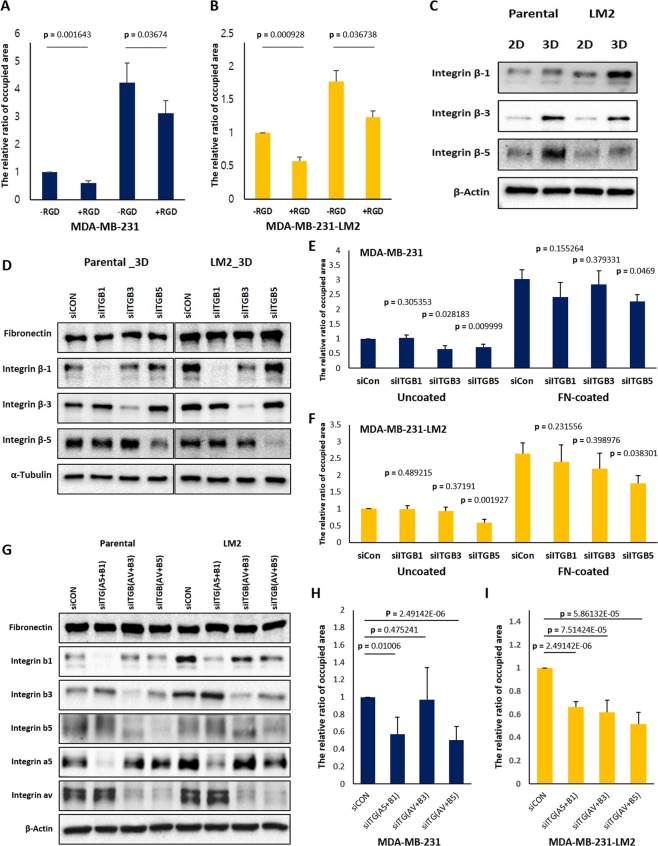
Figure 5.
RGD peptides and integrin β-5 silencing disturb cell attachment from 3D to 2D. (A,B) 500 μM RGD peptides were added into 2D serum-free culture media before cells grown in 3D were transferred on uncoated- or fibronectin coated-2D culture glasses and the cells attached on 2D surfaces were fixed after 2.5 hours. And the areas occupied by the attached cells were measured. (C) Changes in protein levels of each integrin β isoform in different culture conditions were detected, and (D) the protein levels of fibronectin, integrin β-1, integrin β-3, and integrin β-5 of the respective siRNAs treated cells were examined by western blot. The same volume of the set of cell lysates were loaded in different gels to conduct immunoblots for each integrin β isoforms, fibronectin, β-actin, and α-tubulin and the blots were cropped from the gels. (E,F) The siRNA transfected cells were transferred to 2D culture plates uncoated or coated with fibronectin after 3D incubation for 48 hours and the areas covered by attached cells were measured. The mixtures of two different kinds of siRNAs against ITGB1 and ITGB5 and a siRNA against ITGB3 were used for siRNA transfection, respectively. Cells were transfected with siRNAs at concentration of 25 nM in a serial manner, total 50 nM. (G) siRNAs targeting different integrin isoforms were transfected to parental and LM2 cells in three different combinations, and 25 nM siRNA were treated per gene; ITGA5, ITGAV, ITGB1, ITGB3, and ITGB5 (totally 50 nM siRNA for a combination). The mixture of two different kinds of siRNA for ITGA5 and ITGAV were used. (H and I) siRNA transfected cells with each combination were spread on fibronectin coated culture glasses for 2.5 hours after being cultured in 3D suspension condition. And the areas covered by adherent cells were compared with those of control siRNA treated cells. (means ± s.e.m; Student’s t-test).

