Figure 6.
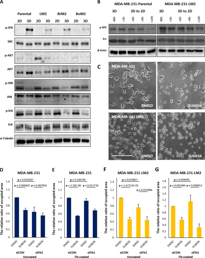
Pharmacological inhibition of SFK reduces the rate of cell-attachment from 3D to 2D. (A) Cells grown in 2D or 3D cultures for 48 hours were harvested and subjected to immunoblot analysis and phosphorylation of signaling proteins were detected. The same volumes of the set of cell lysates were loaded in different gels to carry out immunoblots for p-SFK, Src, Erk, JNK, AKT and α-tubulin, and the blots were cropped from different gels. (B) The changed levels of phosphorylated SFK were observed by western blot. The cells were transferred to 2D culture plate containing serum-free media after 3D suspension culture for 48 hours, and the cell lysates were harvested according to the order of time. The same volume of the set of cell lysates were loaded in different gel to carry out immunoblots for p-SFK, Src and β-actin. (C) SU6656 was added to culture media 30 min before transferring cells from 3D to 2D and the patterns of cell spreading were observed after 2.5 hours. Scale bar: 200 μm. (D–G) Control or fibroenctin down-regulated cells were re-cultivated on uncoated- or fibronectinVI coated-culture glasses after being cultured in 3D suspension condition. Before transferring the cells to 2D, 10 μM of SU6656 was added. The areas covered by attached cells were measured and compared. (means ± s.e.m; Student’s t-test).

