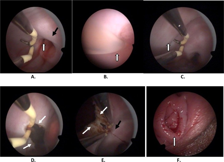
Figure 2.
(A) Vaginoscopic view of cervical canal with spilling of blood (white arrows), left cervix (black arrow). (B) Vaginoscopic view of cervical fistula catheterization (white arrow). (C) Bulging of septum after 5% mannitol injection (white arrow). (D) Hysteroscopic incision at most prominent portion of bulge (white arrows). (E) Upper part of septum has been incised; left cervix visualized (right cervix not visualized due to distance of hysteroscope). (F) An intact hymen after the surgery.

