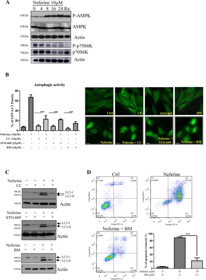
Figure 4.
Role of CaMKKβ-AMPK-mTOR signaling cascade in neferine-mediated autophagy. (A) Neferine activated AMPK-mTOR signaling pathways. HeLa cells were treated with neferine (10 μM) for the indicated time and rapamycin, Ra (300 nM) for 24 h. The cells lysate was then analyzed for p-AMPK, AMPK, p-p70S6K, total p70S6K, and actin respectively. The quantification graphs and full-length blots/gels are presented in Supplementary Figs. S3 and S11 (A), respectively. (B) Inhibitors for AMPK, CaMKKβ and calcium chelator abrogated the neferine-mediated autophagic effect in HeLa cells. HeLa cells were transiently transfected with the EGFP-LC3 plasmid for 24 h and then treated with DMSO (Ctrl), or 10 μM neferine with or without 10 μM of AMPK inhibitor compound C (CC), 25 μM of CaMKKβ inhibitor STO-609 and 10 μM of calcium chelator BAPTA/AM (BM) for 4 h. The cells were then fixed for fluorescence imaging and cells counting. Bar chart represents the quantitation of autophagic cells. (C) Inhibitors for AMPK, CaMKKβ and calcium chelator suppressed the neferine-induced LC3-II conversion in HeLa cells. HeLa cells were treated with DMSO (Ctrl), or 10 μM neferine with or without 10 μM of AMPK inhibitor compound C (CC), 25 μM of CaMKKβ inhibitor STO-609 and 10 μM of calcium chelator BAPTA/AM (BM) for 24 h. Cell lysates were analyzed by western blot for LC3-II conversion (LC3-I, 18 kDa; LC3-II, 16 kDa), and actin respectively. The full-length blots/gels are presented in Supplementary Fig. S11 (B). (D) Calcium chelator, BM inhibits the neferine-induced cell death in HeLa cancer cells. HeLa cells were treated with DMSO (Ctrl), neferine with or without 2.5 μM of BAPTA/AM (BM) for 24 h. The treatment-induced cell death was then measured by flow cytometry analysis after annexin V staining. Bar chart represents the percentage of apoptosis. Data are the means of three independent experiments; error bars, S.D. ***P < 0.001; scale bar, 15 μm.

