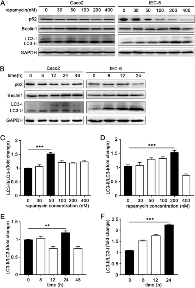
Figure 1.
Induction of autophagy in intestinal epithelial cells. The optimal concentration (A) and time (B) of rapamycin on Caco2 and IEC-6 were respectively detected. Cell lysates were prepared and analyzed by western blotting for the expression of autophagy related proteins LC3, p62 and Beclin1. GAPDH was used as a normalization control. The LC3II/LC3I ratio of Caco2 increased significantly after 24 h incubation with 50 nM rapamycin (C,E), accordingly, 24 h treatment with 200 nM rapamycin could successfully induce autophagy of IEC-6 (D,F). Data was presented as mean ± SD for three independent experiments. **p < 0.01, ***p < 0.001.

