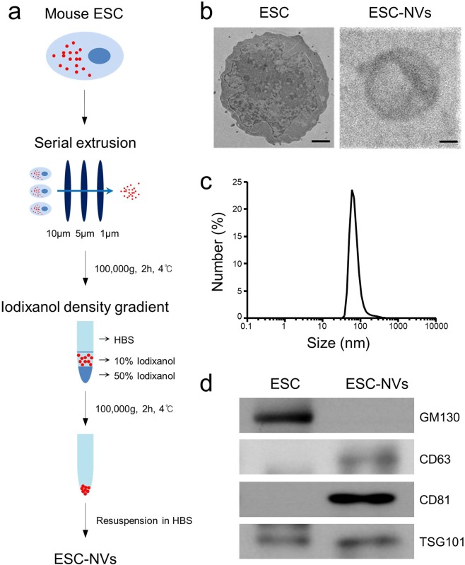
Figure 1.
Preparation and characterization of embryonic stem cell (ESC)-derived extracellular vesicle-mimetic nanovesicles (ESC-NVs). (a) Schematic diagram of the experimental procedure for preparation of ESC-NVs. (b) Representative transmission electron micrograph images of ESC and ESC-NVs. Scale bars = 1000 nm (ESC) or 25 nm (ESC-NVs). (c) Size distribution of ESC-NVs measured by dynamic light scattering analysis. (d) Representative Western blot for a negative marker for NVs (GM130) or positive markers for NVs (CD63, CD81, and TSG101). HBS, HEPES (4-(2-hydroxyethyl)-1-piperazineethanesulfonic acid)-buffered saline.

