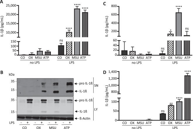
Figure 1.
Calcium oxalate, monosodium urate and ATP induce IL-1β release in LPS-primed murine BMDCs, BMDMs and human PBMCs in vitro. (A,B) Murine bone marrow-derived dendritic cells (BMDCs), (C) murine bone marrow-derived macrophages and (D) human peripheral blood mononuclear cells were primed with LPS (100 ng/ml) for 3 hours before either calcium oxalate crystals (100 µg/ml) (OX), monosodium urate crystals (300 µg/ml) (MSU) or ATP (5 mM) or no further stimulus as control (CO) were added for an additional 6 hours. Culture supernatants (SN) were collected and concentrations of IL-1ß were measured using ELISA. (A,C,D) In addition, supernatants and whole cell lysates (LY) from stimulated murine BMDCs were analyzed by western blotting. (B) All three stimuli induced IL-1ß release in all three cell types. Data are presented as mean ± SD of a representative experiment of a total of nine each performed with triplicate biological samples. Statistical analysis was performed using two-way ANOVA and post hoc analysis. ****P < 0.0001; **P < 0.01; *P < 0.05; ns, not significant compared with control treatment (without LPS). To improve the clarity of the presented western blots, blots are displayed in a cropped version. Full-length gels are presented in Supplementary Fig. 1.

