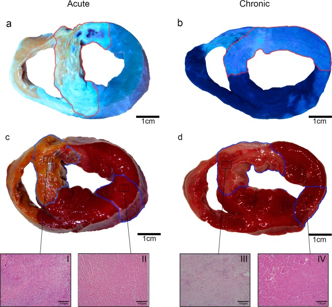
Figure 5.
Macroscopic heart sections from the acute (left panels) and chronic (right panels) myocardial infarction groups. Upper panels (a,b): Area at risk corresponds to tissue stained with thioflavin-S (light blue). Lower panels (c,d): The infarcted tissue corresponds to 2,3,5-Triphenyltetrazolium non-stained myocardium. Samples were obtained from the acute (c: I-infarct, II-remote) and chronic infarct models (d: III-infarct and IV-remote) and stained with haematoxylin-eosin.

