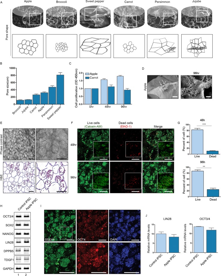
Figure 2.
Induced pluripotent stem cells (iPSCs) cultivated in apple-derived scaffolds: (A) Drawings of various plant scaffolds showing shape and pore sizes; (B–D) Pore sizes, cell proliferation assay (CCK-8), and scanning electron microscopic images of human induced pluripotent stem cells (hiPSCs) seeded in apple scaffolds; (E) Phase contrast (top) and haematoxylin & eosin-stained (bottom) images of hiPSCs seeded onto apple scaffolds (original magnifications: 100x and 200x); (F) Views of live and dead cells in cultures after 48 h (top panels) and 96 h (bottom panels); (G) Proportion of the live and dead cells in cultures after 48 h and 96 h; (H) mRNA expression levels of iPSC markers in control iPSCs and iPSCs cultured in apple scaffolding; (I) Immunofluorescence staining of iPSC markers in iPSCs cultured in apple scaffolding; and (J) mRNA expression levels of LIN28 and OCT3/4 in control iPSCs and iPSCs cultured in apple scaffolding (*p < 0.01; **p < 0.005; and ***p < 0.001).

