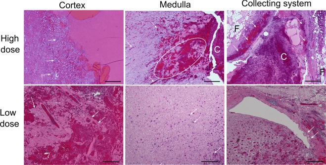
Figure 3.
Different renal tissues - cortex, medulla and collecting system – showed differential sensitivities to BH ablation. Histological slides of the volumetric lesions stained with H&E were taken from the tissues subjected to the same BH dose, i.e. number of pulses delivered per focal spot. At the higher dose of 15–30 pulses per focal spot renal cortex is fully liquefied with no debris larger than 20 microns observed. White arrows indicate intact glomeruli adjacent to the liquefied region. In medulla, areas of disrupted, but not completely disintegrated connective tissue are observed (dashed circle), as well as blood within tubules (white arrows) that appears to flow towards collecting system and form a clot (C). Collecting system containing the clot has minimal observable damage in terms of bruising and sloughing of the urothelium (white arrows); the fat layer (F) appears intact. At low dose (5–10 pulses/spot) less tissue damage is noted within all three tissues. In the cortex, connective tissue fragments and structures with adjacent cells are observed within the lesion (white arrows). In medulla, only blood within the tubules is observed (white arrows), and very minor urothelium sloughing is seen in the collecting system (white arrows). The scale bar represents 500 microns.

