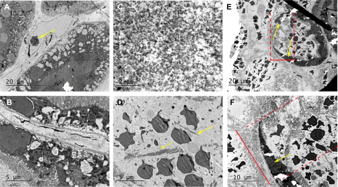
Figure 4.
Transmission electron microscopy (TEM) of renal cortex ablated with BH. (A,B) TEM images of intact renal tissue adjacent to the BH lesion demonstrating proximal renal tubules and small caliber blood vessel with lymphocyte (arrow). (C,D) BH lesions containing a slurry of homogenized cellular debris <0.1 μm in size (C), occasional loose collagen fibers (arrows), and echinocytes (D). (E,F) At the lesion border, the transition zone width was 20 microns from fully intact to fully destroyed tissue (top red bar/dashed lines). Within the zone, the cells (arrows) are significantly damaged, without vili but containing some organelles (bottom).

