Figure 3.
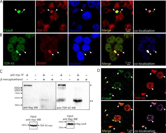
TDP-43 co-localizes with f-LeuR and endogenous RGNEF within micronuclei and interacts in vitro and co-localizes in vivo with f-LeuR. (A,B) Representative confocal images of HEK293T cells showing co-localization (white arrows) of endogenous TDP-43 with f-LeuR (A) or endogenous RGNEF (B) within micronuclei after cellular metabolic stress using lactate. (C) IP of TDP-43-myc after crosslinking using DTSSP on protein lysate from HEK293T cells expressing f-LeuR and TDP-43-myc. WB was performed for detecting flag and then TDP-43 after stripping. Input controls are showed. β-mercaptoethanol was used to dissociate the crosslinked complex (* and ** mark electrophoretic shifts of approx. 440 kDa and 60 kDa, respectively). (D) Schematic of scAAV-9-LeuR virus and representative confocal images showing the extensive co-localization (white arrows) between LeuR and TDP-43 in brain of rats 4 weeks after the injection with the virus that express flag-LeuR in neurons under SYN1 promoter (yellow arrows indicate granular L-rich that doesn’t co-localize with TDP-43). The proteins were detected using goat anti-flag and rabbit anti-TDP-43 antibodies.

