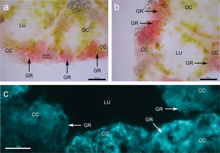
Figure 2.
Zinc distribution in midgut gland sections of zinc-exposed Arion vulgaris with colour dithizone staining (a,b) and fluorescent toluenesulfonamidoquinoline (TSQ) staining (c). Midgut gland cross sections showing lumen (LU) surrounded by digestive cells (DC) and calcium cells (CC) containing calcium granules (GR). Zinc stained by dithizone (red colour) (a,b) is exclusively allocated in calcium cells. Fluorescent staining by TSQ localize zinc (bright blue colour) (c) mainly in cytoplasm of calcium cells and on the outer edge of calcium granules (c - GR). The bar corresponds to a size of 50 µm.

