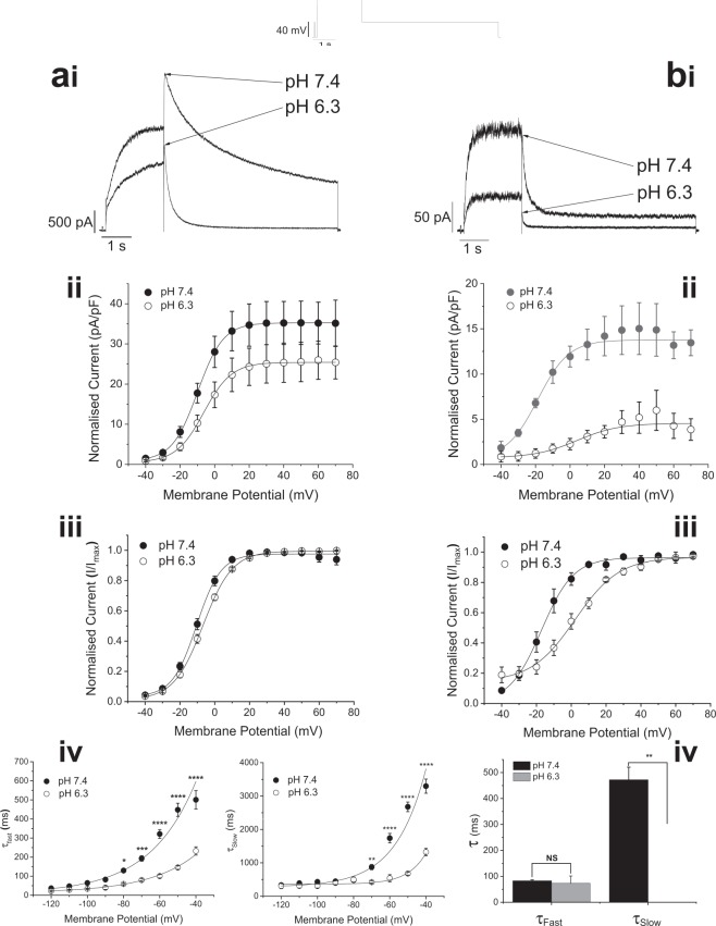
Figure 1.
hERG1a- and 1b-mediated current is inhibited by extracellular acidosis. (ai) HERG1a-mediated current evoked by the protocol in inset is reduced in amplitude and current deactivation is accelerated by addition of extracellular solution with a pH of 6.3. (aii and iii) Extracellular acidification causes a shift in voltage-dependence of activation of hERG1a-mediated current, with V0.5 of −10.0 ± 1.2 mV (k = 6.8 ± 0.2 mV) at pH 7.4 (n = 15 cells) shifted to a V0.5 of −7.0 ± 2.8 mV (k = 8.3 ± 1.1 mV) at pH 6.3 (n = 8 cells). (aii) Shows mean current-voltage (I–V) relations for IhERG tails at pH 7.4 and 6.3, expressed in pA/pF, whilst in (aiii) currents following each test potential were normalized to the maximum current (Imax) elicited by the protocol. (aiv) Graphs showing acceleration by acidic pHe of the two time -course components of hERG1a-mediated current with hyperpolarization. Data are shown as mean ± S.E.M for the fast time constant (τFast) (left) at pH 7.4 (•)(n = 15 cells) and 6.3 (○)(n = 8 cells) and slow time constant (τSlow) (right) of deactivation at pH 7.4 (•)(n = 15 cells) and 6.3 (○)(n = 8 cells). The fast component of current deactivation changed e-fold in 26 mV. Application of pH 6.3 accelerated the fast time component without affecting apparent voltage dependence. In contrast, the slow component of deactivation changed e-fold in 20 mV, with pH 6.3 solution increasing the rate of this component and reducing the apparent voltage dependence to e-fold in 30 mV. ‘*’, ‘**’, ‘***’, and ‘****’ denote statistical significance of P < 0.05, P < 0.01, P < 0.001 and P < 0.0001 respectively (2-way ANOVA with Bonferroni post-test). (bi) hERG1b-mediated current (same protocol as hERG1a) was reduced in amplitude and current deactivation is accelerated by addition of extracellular solution with a pH of 6.3. (bii and iii) The voltage dependence of activation of hERG1b-mediated current was significantly positively shifted, with the V0.5 value changing from −16.2 ± 2.8 mV (k = 7.6 ± 0.6 mV) at pH 7.4 (n = 7 cells) to a V0.5 of +2.2 ± 1.1 mV (k = 12.6 ± 1.0 mV) at pH 6.3 (n = 7 cells). (bii) Shows mean current-voltage (I–V) relations for IhERG tails at pH 7.4 and 6.3, expressed in pA/pF, whilst in (biii) currents following each test potential were normalized to the maximum current (Imax) elicited by the protocol. (biv) The time-course of the deactivation of hERG1b-mediated current was accelerated by reduction of pHe. The bar chart shows mean ± SEM data for the fast (τFast) and slow (τSlow) time constant components of hERG1b deactivation measured upon repolarization to −40 mV. Addition of pH 6.3 solution did not affect τFast (P = 0.7469; paired t-test), but significantly accelerated τslow (P = 0.0074; paired t-test).

