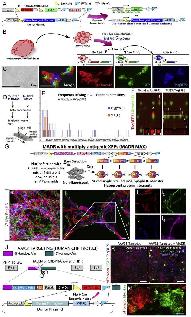
Figure 1: MADR in mTmG mouse or human lines generates genetic reporter-defined populations in vitro.
A) Flp-Cre vector catalyzes either Cre-mediated excision or dRMCE on Rosa26mTmG allele in the presence of a MADR donor vector, resulting in two distinct recombinant products.
B) Nucleofection of heterozygous Rosa26WT/mTmG mNSCs result in three possible lineages: tdTomato+, EGFP+, and TagBFP2+.
C) Live imaging of representative cells with non-overlapping fluorescent colors. Scale bars, 100μm
D) Schematic of cell preparation for single-cell western blot.
E) Frequency of fluorescence intensities comparing MADR and PiggyBac transgenic cells.
F) Representative examples of single-cell western blots for PiggyBac and MADR groups. (Note that this is not a pure population and so some cells express the Histone H3 loading control protein but no TagBFP2. Also, many lanes are empty as is typical for this assay).
G) MADR-compatible TRE-smFP plasmids for MADR MAX.
H) Dox induces efficient smFP expression allowing for orthogonal imaging of 4 independent reporters in vitro. Scale bar, 100μm
I) High magnification confocal z-section demonstrates that each cell expresses a single smFP reporter. Scale bar, 10μm
J) Schematic of AAVS1 locus targeting for HUMAN MADR by TALEN or CRISPR/Cas9
K) HEK293T cells containing AAVS1-targeted MADR recipient site expressing tdTomato and TagBFP2-V5-nls Scale bar, 100μm
L) MADR-HEK293T cells transfected with MADR pDonor smFP-myc (Bright) or TagBFP-3XFlag showing GFP or BFP autofluorescence among non-inserted tdTomato+ cells. Scale bar, 100μm
M) High mag image of cells from L exhibiting tdTomato and smFP-myc in a mutually exclusive manner. Scale bar, 10μm

