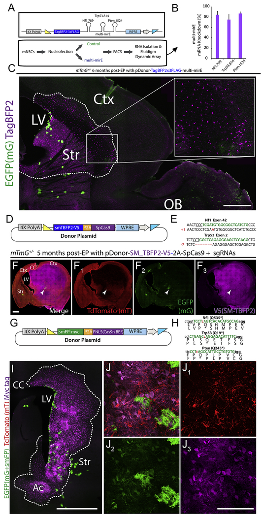
Figure 3: Loss-of-function manipulations using MADR transgenesis.
A) Donor construct for miR-E shRNAs against Nf1, Pten, and Trp53 tied to TagBFP2 reporter
B) Validation of knockdown efficacy of multi-miR-E function by qPCR.
C) 6-month-old mouse sagittal section showing a hyperplasia of TagBFP2+ cells but no tumor. Scale bar, 1mm
D) Plasmid for MADR of a TagBFP2-V5 reporter protein and SpCas9
E) Sequencing of TdTomato-/EGFP- glioma cells exhibit InDels in Nf1 and Trp53.
F) MADR insertion of TagBFP2-V5 reporter and Cas9 with co-EPed PCR-derived sgRNAs yields high grade glioma observable through labeling of 3 genetic reporter-defined populations in a coronal section of both hemispheres. Scale bar, 1000μm
G) Plasmid for MADR of an smFP-myc reporter protein and FNLS Cas9n base editor.
H) sgRNA-targeting sites (green letters) induce C->T base conversion (red lowercase ‘c’ are targeted) to produce premature stop codons in Nf1, Trp53, and Pten.
I) MADR insertion of myc reporter and FNLS Cas9n with co-EPed PCR-derived sgRNAs yields observable expansion of OPC progenitors at two months post-EP through labeling of three genetic reporter-defined populations in a coronal section. Scale bar, 1000μm
J) High magnification tdTomato (1), EGFP (2), and Myc tag (3) image showing myc+ populations. Scale bar, 100μm

