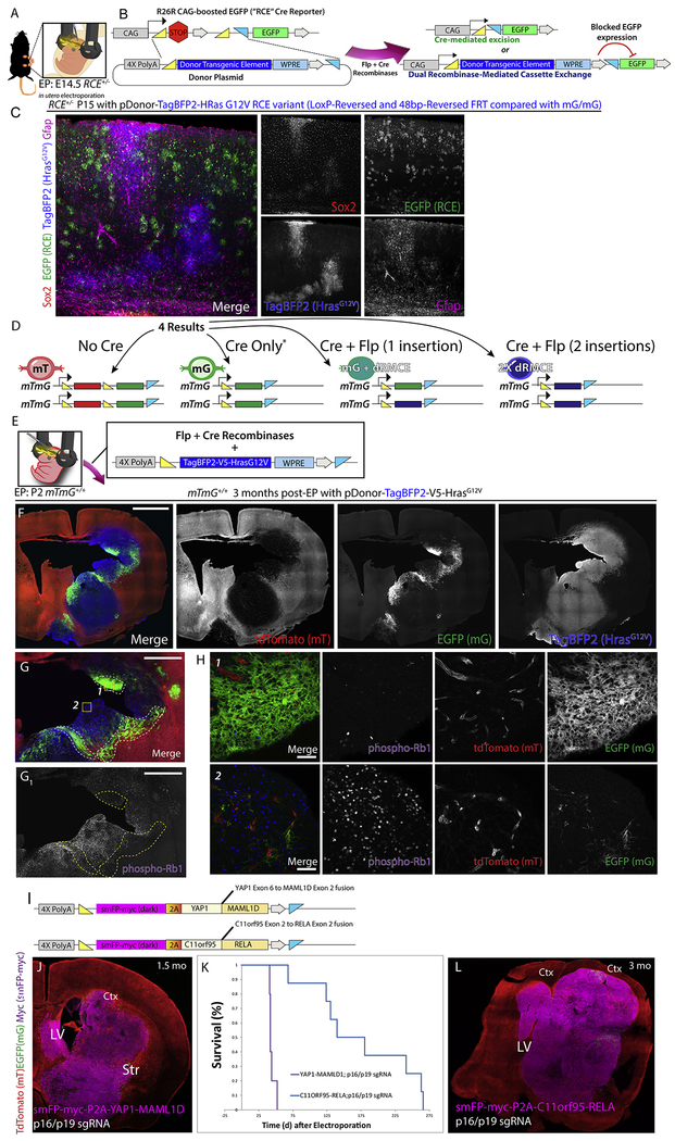
Figure 4: Generation of somatic glioma using in vivo MADR with HrasG12V indicates dosage effects of this oncogene and human oncofusion proteins generate ependymal tumors.
A-B) Schematic for in utero EP of MADR into E14.5 RCE +/− dams
C) In utero EP in RCE mice with HrasG12V oncogene produces mosaic patches of TagBFP+ astrocytes Rosa26HrasG12 but not evidence of invasive glioma
D) Schematic of possible outcomes after MADR in homozygous mt/mg recipient mice
E) P2 EP of homozygous mt/mg mice with TagBFP2-HrasG12V oncogene
F) Postnatal EP in homozygous Rosa26mTmG P2 pups with HrasG12V oncogene produces two different tumor types (Blue-only Rosa26HrasG12V×2 and blue-and-green Rosa26HrasG12V×1) Scale bars: 2mm
G) Representative tumor formation in homozygous mTmG 3 months post-EP. Blue-only Rosa26HrasG12V×2 cells occupy a larger section of the tumor than blue-and-green Rosa26HrasG12V×1, correlating with phosphor-Rb1 protein expression. Scale bars: 1mm
H) Zoom-in images of regions 1 and 2 from G show phosphorylated-Rb1 expression correlates largely with blue-only cells. Scale bars: 50μm
I) Plasmid schematics for expression of ependymoma-associated fusion proteins
J) Stitch of YAP1-MAML1D; p16/p19 Cas9 targeting induced ependymoma-like tumor.
K) Survival analysis of Ependymoma MADR model mice
L) Ependymoma-like tumor in a 3-month-old C11orf95-RELA; p16/p19 Cas9-targeted mouse

