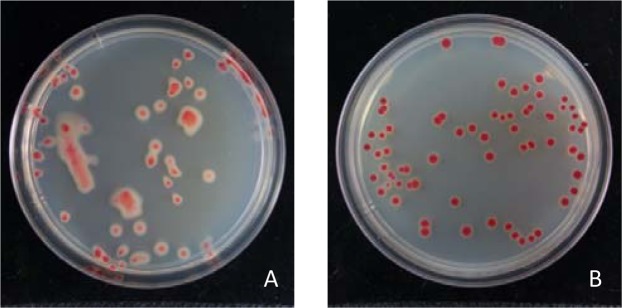
Figure 2.
Colony morphology of virulent (A) and avirulent (B) Ralstonia solanacearum strains on TTC medium. The isolated R. solanacearum from the test samples showed two different colony morphologies on TTC medium. The colony of virulent R. solanacearum was irregular, highly mobile, humid and displayed a pink spot in the middle of the colony and a large white edge (A); while the colony of avirulent R. solanacearum was round, immobile, dry and displayed a dark red spot in the middle of colony and a narrow or no white edge (B).

