Figure 2.
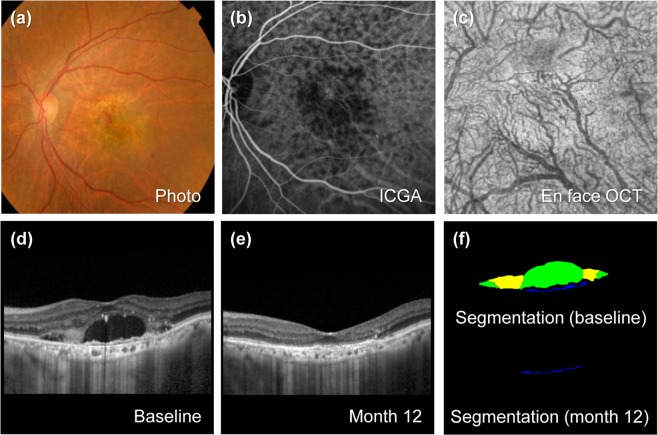
Representative case of typical neovascular age-related macular degeneration (nAMD) without pachychoroid. The patient is an 80-year-old woman with a baseline visual acuity of 20/100. For 45.4 months of follow-up, the eye was treated with 9 intravitreal injections of ranibizumab. (a) Baseline fundus photography shows subretinal exudation and hemorrhage change at macula. (b) Baseline indocyanine green angiography (ICGA) shows choroidal neovascularization (CNV) without polyps. (c) En face optical coherence tomography (En face OCT) scan at the final visit. Choroidal vessels spanning the macular area are not dilated. (d) Enhanced depth imaging optical coherence (EDI OCT) at baseline. The subfoveal choroidal thickness (SFCT) was thin as 91 µm. (e) EDI OCT scan 12 months from baseline. Visual acuity improved to 20/30 at 12 months. At the final visit, visual acuity improved to 20/40. (f) Upper panel shows predicted lesion corresponding baseline OCT B-scan by a convolutional neural network (CNN) (upper). Subretinal fluid (SRF, green color), subretinal hyperreflective material (SHRM, yellow color), and pigment epithelial detachment (PED, blue color) are noted. Each component could be quantified by converting the pixel counts into mm2, and the areas of SRF, SHRM and PED are 0.388, 0.143, and 0.027 mm2, respectively. Lower panel shows PED predicted from OCT B-scan at 12 months by CNN, and the area is 0.017 mm2.

