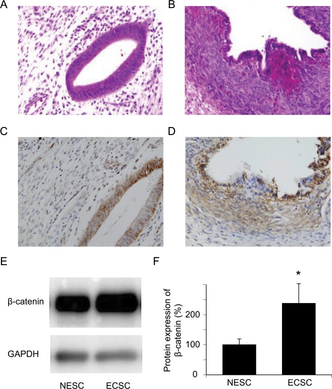
Figure 1.
Expression of β-catenin is upregulated in endometriosis. (A) HE staining of a normal endometrium. An endometrial gland and endometrial stromal cells are shown. (B) HE staining of endometriosis. (C) A representative image of immunohistochemical staining of a normal endometrium with anti-human β-catenin. (D) A representative image of immunohistochemical staining of endometriosis with anti-human β-catenin. (E,F) Significant upregulation of β-catenin protein expression in ECSC compared with NESC is shown by western blotting. n = 5, *p < 0.01, Student’s t-test. Error bars represent standard deviation (SD). Uncropped images are shown in Fig. S5.

