Figure 1.
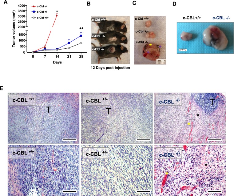
Reduced c-Cbl activity enhances tumor growth and immune infiltrates. (A) Average of tumor volumes from six mice injected subcutaneously with MC38 cells in c-Cbl+/+ and c-Cbl+/− groups and five in the c-Cbl−/− group. All the c-Cbl−/− mice had to be euthanized by 10–12 days with rapid development of the xenograft along with ulceration on the tumor. Student’s t-test was performed. Error bars = SEM. *p < 0.0001 corresponds to growth of tumors in c-Cbl−/− mice compared to both c-Cbl+/− and c-Cbl+/+ groups. **p = 0.003 compares the group tumor growth c-between Cbl+/− and c-Cbl−/− group. (B) A representative mouse from three groups is shown at 12 days post-injection. (C) Excised xenografts are shown. Blue asterisk corresponds to the areas of necrosis and liquefaction (soft by palpation) compared to other areas of xenografts and yellow asterisk demarcates areas of ulceration. (D) Excised MC38 xenografts are shown. Representative images of a pair of c-Cbl+/+ and c-Cbl+/− mice are shown. (E) H&E stain of xenografts from different groups of mice are shown at 40X and 100X magnification. Xenografts from c-Cbl+/+ and c-Cbl+/− mice showed tumor cells (T) without evidence of acellular areas with fragmented nuclei suggestive of necrosis. Xenografts from c-Cbl−/− mice showed acellular areas suggestive of necrosis (marked with a black asterisk) and several areas of pools of RBCs suggestive of hemorrhage (marked with a yellow asterisk). Scale bar = 25 micron (40×) and 50 micron (100×).

