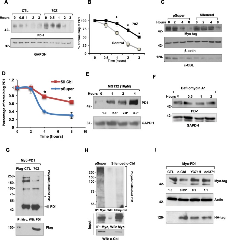
Figure 7.
c-Cbl destabilizes PD-1 and targets it for ubiquitination and proteasomal degradation. (A) RAW264.7 expressing control (CTL) and 70Z-c-Cbl were treated with Cycloheximide (300 μM) for the indicated time. Representative immunoblots from four experiments are shown. (B) Densitometry of PD-1 normalized to GAPDH and represented as the percentage of PD-1 at time = 0. Time to reach 50% of PD-1 was considered as its half-life. Average of four experiments is shown. Error bars = SEM. Student’s t-test was used. *p = 0.03 and **p = 0.01. (C) Lysates from HEK-293T cells stably expressing control (pSuper) and c-Cbl shRNA (Silenced) vectors were transfected with Myc-PD-1 and treated with Cycloheximide (100 μM) for the indicated time. The amount of remaining PD-1 was detected by Western blotting. Equal amounts of lysates were probed separately to confirm c-Cbl silencing. A representative figure from four independent experiments is shown. (D) Densitometry of normalized PD-1 bands performed using ImageJ represented as the percentage of PD-1 at time 0 is shown. The time to reach 50% of initial PD-1 was considered as the half-life of PD-1. An average of four experiments is shown. Error bars = SD. Student’s t-test was performed. *p = 0.001 and **p = 0.003. (E) RAW264.7 cells treated with MG132 for indicated amount of time. Representative immunoblots from four independent experiments are shown. Student’s t-test with Bonferroni correction was used. Compared to time = 0, *p = 0.04 at 1 hour, p = 0.001 at 2 and 4 hours. (F) Raw264.7 cells treated with 200 nM of Baflomycin A1 for the indicated time points. The lysates were probed as shown. A representative of two experiments is shown. (G) HEK 293T cells expressing control (Flag-CTL) and 70Z-c-Cbl were pre-treated with 10 µM MG132 overnight and immunoprecipitated using PD-1 antibody. The eluents were probed with PD-1. 5% of lysates were probed separately with Flag-tag. Representative immunoblots from three experiments are shown. (H) HEK293T cells stably expressing shRNA of c-Cbl (Silenced c-Cbl) and control (pSuper) and transfected with Myc-tagged PD-1 were immunoprecipitated using Myc antibody. Eluents were probed with anti-ubiquitin antibody. 5% of lysates were probed for c-Cbl. Representative immunoblots from three experiments are shown. (I) HEK293T co-expressing Myc-tagged PD-1 and HA-tagged c-Cbl or control (CTL) constructs were probed and normalized to actin. Equal amounts of lysates were probed for HA-tag. Representative immunoblots from three independent experiments. 2-way ANOVA with multiple comparison and Student’s t-test were performed. Compared to CTL, *p = 0.02 for c-Cbl.

