Figure 5.
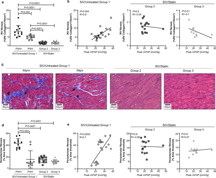
Effect of statins on SIV-associated fibrosis in the heart and pulmonary arteries. (a) Quantification of right ventricle (RV) collagen deposition quantified from Masson’s trichrome-stained heart sections. (b) Correlation analysis between peak mPAP and RV fibrosis (%RV Collagen/170 mm2) in SIV/Untreated animals (left), and statin-treated cohorts (center, right). (c) Representative images of Masson’s trichrome-stained right ventricle sections. (d) Quantification of pulmonary periarteriolar collagen deposition quantified from Picro-Sirius Red-stained lung sections. (e) Correlation analysis between peak mPAP and periarteriolar fibrosis (%Area threshold) in SIV/Untreated animals (left), and statin-treated cohorts (center, right). (a,d) Mann-Whitney U test was used for statistical analysis. Data represents the mean ± SD. (b,e) Spearman correlation analysis; R, Spearman coefficient.

