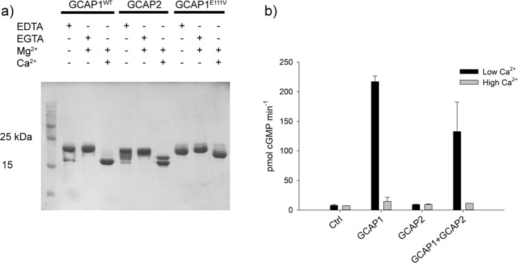
Figure 1.
Purity of recombinant proteins, Ca2+-induced gel shifts and GC1 enzymatic assays. (a) SDS-PAGE of WT/E111V GCAP1 and GCAP2 in the presence of 5 mM EDTA, 4 mM EGTA + 1.4 mM Mg2+ and 1 mM Mg2+ + 4 mM Ca2+. Twenty micromolar WT-GCAP1, E111V-GCAP1 and WT-GCAP2 were incubated for 10 min at 30 °C in the presence or absence of ions and loaded in a 15% SDS-PAGE. Gel was Coomassie blue-stained. (b) GC1 enzymatic assays in the presence of 10 μM GCAP1, 10 μM GCAP2 and equal amounts of both (5 μM GCAP1 + 5 μM GCAP2) in the presence of less than 19 nM Ca2+ (low Ca2+) or ~ 30 μM Ca2+ (high Ca2+). Each bar represents the average of three replicas ± standard deviations (n = 3). Full-length gels are reported in Supplementary Fig. S1.

