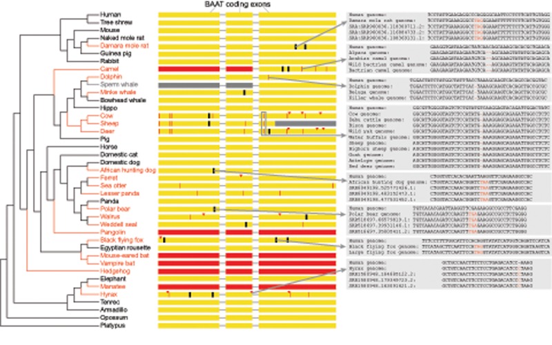
Fig. 1.
—Repeated losses of BAAT in placental mammals. A phylogenetic tree shows the species that have gene-inactivating mutations in BAAT (red font) together with a subset of species that have an intact BAAT gene (black font). The three coding exons of BAAT are shown as yellow boxes together with the position of inactivating mutations. Red arrowheads represent frameshifting insertions, red vertical lines represent frameshifting deletions, black vertical lines represent premature stop codons, and red boxes represent exon deletions. Gray insets illustrate that all shown inactivating mutations are validated either by unassembled sequencing reads or by the presence of the same mutation in the genomes of related species. The downstream parts of exon 3 in sheep (gray color) overlap an assembly gap; however, sheep shares other inactivating mutations with related species, showing that BAAT is lost in the sheep. The status of BAAT in the sperm whale is uncertain as the genomic region around exons 1 and 2 is not properly assembled (scaffolds end up- and downstream of these exons and there is no colinear alignment). Based on inactivating mutations that are shared between related species, we infer as many as 18 independent losses of BAAT have occurred in placental mammals.

