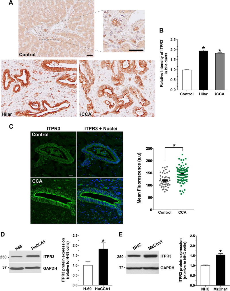
Figure 1. ITPR3 staining is increased in cholangiocytes of patients with hilar and intrahepatic cholangiocarcinomas and in cholangiocarcinoma cell lines.

(A) Representative immunohistochemical (IHC) staining of ITPR3 in liver biopsy specimens from controls and patients with hilar cholangiocarcinoma and intrahepatic cholangiocarcinoma (iCCA) shows that ITPR3 staining is more intense in liver samples of CCA patients. Scale bars: 50 μm. (B) Quantitative analysis of the intensity of ITPR3 staining in the IHC images. Images are representative of what was observed in 3–7 patients, using 4–5 images per patient, *P < 0.0001. (C) Representative confocal immunofluorescence images of ITPR3 (left) and quantitative analysis of the intensity of ITPR3 staining in the images (right) of liver biopsy specimens from controls and CCA patients. ITPR3 expression (green) is detected only in bile ducts. Nuclei were labeled with a nuclear marker (blue). Labelling of ITPR3 in cholangiocytes in CCA patients (n=97 ducts from 7 patients) is greater than in controls (n=47 ducts from 7 patients; *P < 0.0001). Representative immunoblotting (top panel) and relative protein expression (bottom panel) of ITPR3 (D) in H69 and HuCCA1 cells and (E) in NHC and MzCha1 cells show that ITPR3 is increased in cholangiocarcinoma cell lines. *P < 0.05 (H69 versus HuCCA1) and *P < 0.001 (NHC versus MzCha1) (n=6).
