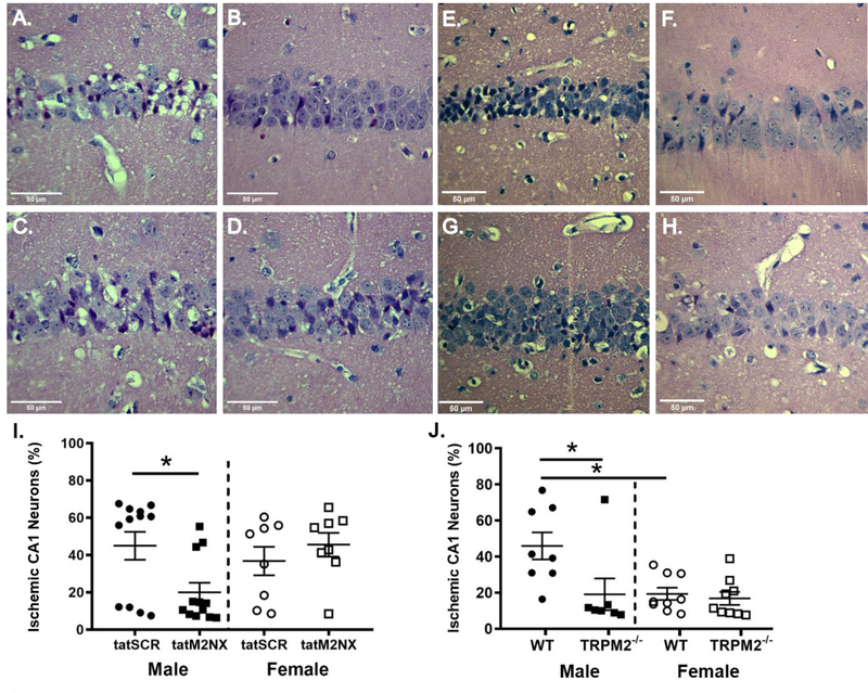
Figure 1. Acute tatM2Nx reduces neuronal injury in males.
Representative photomicrographs of hippocampal CA1 neurons from male mice injected with 2o mg/kg scrambled control peptide; tat-SCR male (A) and female (C) or 2o mg/kg tatM2NX male (B) and female (D) 3o minutes after resuscitation. (I) Quantification of ischemic CA1 neurons 3 days after recovery from CA/CPR in mice treated with tatSCR vs tatM2NX. Representative photomicrographs of hippocampal CA1 neurons from WT male (E) and female (G) or TRPM2−/− male (F) and female (H) mice 3 days after recovery from CA/CPR. J) Quantification of ischemic CA1 neurons 3 days after recovery from CA/CPR. *p < 0.05. Data presented as mean±SEM.

