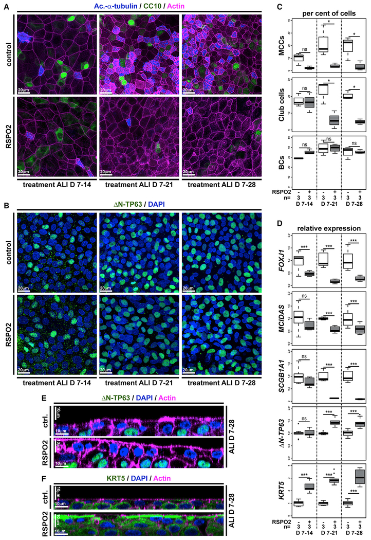
Figure 4. Wnt/β-Catenin Signaling Inhibits Differentiation and Promotes Stemness in Human BCs.
(A–F) Human immortalized BC (BCIs) kept in air-liquid interface (ALI) culture for up to 4 weeks. Human recombinant R-spondin2 (RSPO2) was used to activate Wnt/β-catenin signaling starting at ALI day 7 (D7). n = 3 cultures per time point and treatment.
(A) Confocal imaging of specimens stained for Ac.-α-tubulin (MCCs, blue), CC10 (Club cells, green), and Actin (cell membranes, magenta) show reduced MCC and Club cell numbers in RSPO2-treated cultures.
(B) RSPO2 does not reduce the number of ΔN-TP63+ (green) cells. Nuclei (DAPI, blue).
(C) Quantification from (A) and (B). Mann Whitney test, not significant, ns (p > 0.05); *p ≤ 0.05; **p ≤ 0.01; ***p ≤ 0.001.
(D) Quantitative real-time PCR. Expression levels are depicted relative to stage controls. RSPO2 reduces expression of MCC (FOXJ1, MCIDAS) and Club cell (SCGB1A1) markers but increases expression of BC markers (ΔN-TP63, KRT5). Student’s t test, not significant, ns (p > 0.05); *p ≤ 0.05; **p ≤ 0.01; ***p ≤ 0.001.
(E and F) Optical orthogonal sections of confocal images. RSPO2-treated cultures display increased epithelial thickness and cells staining for BC markers ΔN-TP63 (green, in E) and KRT5 (green, in F).
Related to Figure S4.

