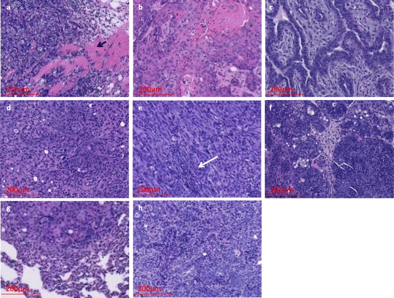
Fig. 2.
Histology of Pik3caH1047R;Trp53R270H;MMTV-Cre primary mammary tumors and metastasis. a Invasive poorly differentiated adenocarcinoma (arrow indicates muscle fibers), b adenosquamous carcinoma, c fibroadenoma, d sarcomatoid adenocarcinoma, e sarcomatoid (spindle cell) carcinoma with lymphocyte infiltration (white arrow indicates site of lymphocyte infiltration), f poorly differentiated carcinoma, g metastatic lung carcinoma (from the same animal for which primary tumor is shown in panel d), and h metastatic lymph node (from the same animal for which primary tumor is shown in panel e)

