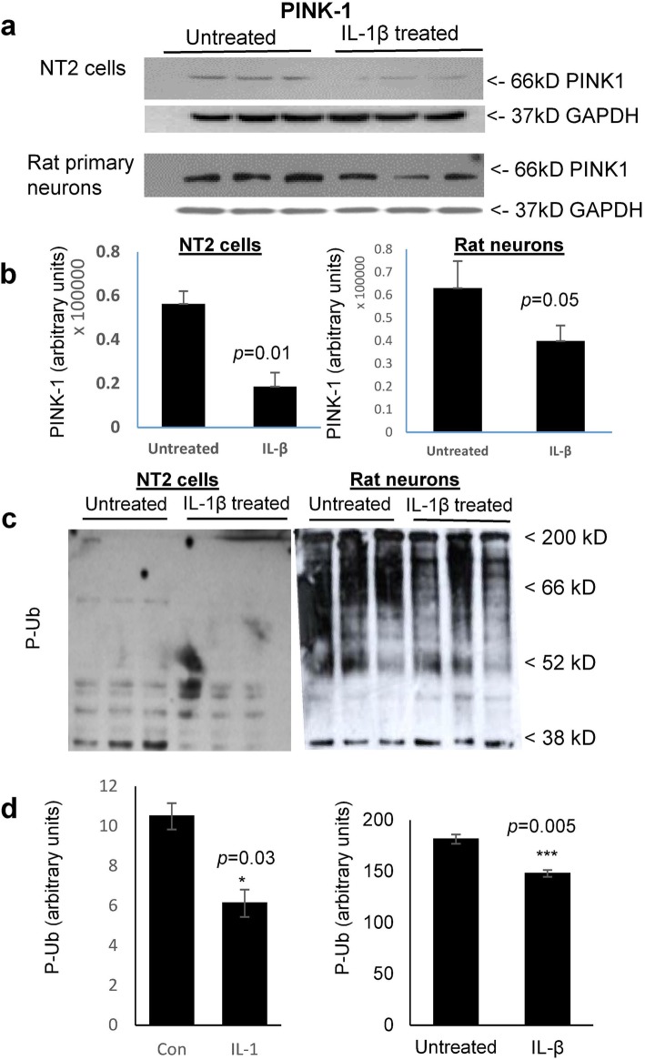
Fig. 2.
IL-1β treatment of both NT2 cells and rat primary neurons decreased the steady-state levels of PINK1 and of Ser65-phosphorylated ubiquitin (P-Ub); conversely, IL-1β increased the levels of Ser65-phosphorylated parkin (P-parkin). a Representative western immunoblots depicting PINK1 levels in IL-1β treated vs untreated NT2 and rat primary neuronal cell cultures, representing 6 independent experiments. b Quantification of PINK1 levels in both NT2 cells (p = 0.01), and primary rat neurons (p = 0.05). c Representative western immunoblots of IL-1β treated vs untreated NT2 and rat primary neuronal cell cultures (n = 6 and 3 technical repeats, respectively). d Quantification of P-Ub levels in both NT2 (p = 0.03) and rat primary neurons (p = 0.005)

