Fig. 4.
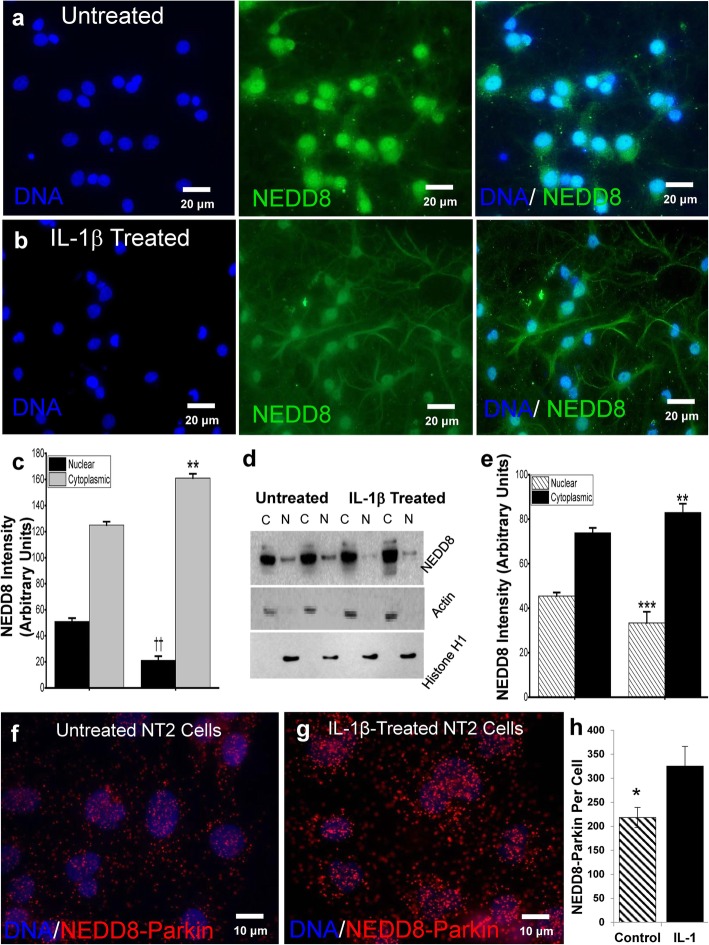
IL-1β treatment of primary neuronal cultures leads to NEDD8 translocation. Fluorescent immunocytochemistry of representative cultures of untreated (a) and IL-1β-treated (b) primary rat neurons (n = 6 each) depict greater translocation of NEDD8 (green) from nucleus (blue) to cytoplasm when IL-1β is present, **p < 0.01, ††p < 0.01 (c). Quantification of western immunoblots (d) of nuclear (histone) and cytoplasmic (actin) cell fractions from untreated and IL-1β-treated cultures (n = 6 separate experiments) demonstrates the susceptibility of NEDD8 to undergo nuclear to cytoplasmic translocation in the presence of excess IL-1β, **p < 0.01, ***p < 0.01 (e). Quantification of NEDD8/parkin colocation (≤ 40 nm) in untreated NT2 cells (f) compared with IL-1β-treated NT2 cells (g) provides evidence of IL-1β-induced interaction between of parkin and NEDD8 in NT2 cells, two-tailed t test, *p < 0.05 (h). NEDD8-parkin proximity (red dots) was quantified with FIJI (ImageJ) Particle Analysis

