Figure 1.
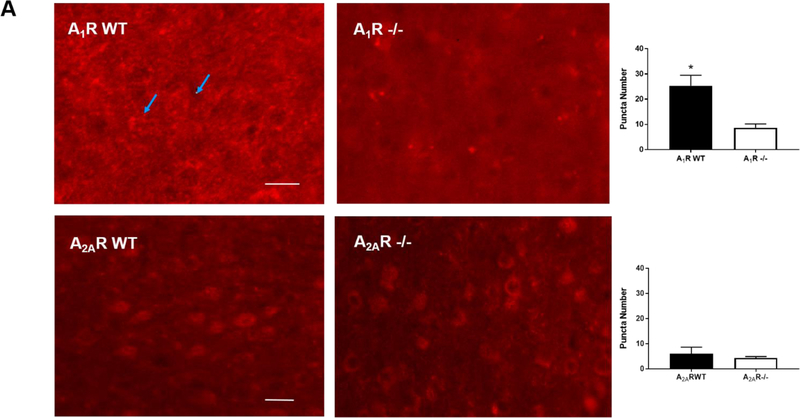
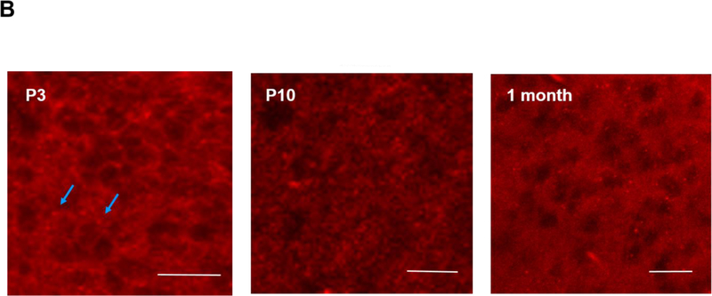
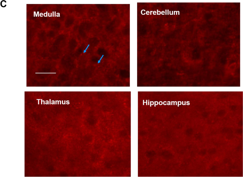
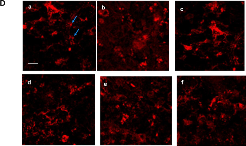
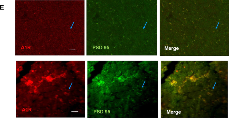
The brains of neonatal mice and preterm infants expressed A1R receptors. (A) Immunostaining results showed the specificity of A1R antibody but not A2AR antibody. Using the tested A1R antibody, A1R immunostaining showed a punctate distribution of this receptor in the cortex of wild type adult mice, but not in that of A1R deficient mice (25.33±2.4 and 8.67±0.88 puncta, respectively) (top). In contrast, several antibodies against A2AR showed non-specific immunoreactivity in the nucleus and the cytoplasm in both wild type and A2AR deficient mice (bottom). No punctate pattern was observed (6±1.53 puncta for WT and 4.33±0.3 puncta for A2AR knockout). Representative images are shown here. The quantitative results are shown on the right. Blue arrows indicate puncta. Scale bar, 10 μm. (B) The punctate pattern of A1R could be detected in the cortex of P3 pups. The A1R puncta appeared more discrete at a lower magnification as the animals mature (10 day-old and 1 month-old pups). (C) A1R distributed in a punctate pattern in medulla, cerebellum, thalamus and hippocampus. The puncta were more prominent in medulla and cerebellum. Scale bar, 20 μm. (D) Immunostaining showed a punctate pattern of A1R in the cortex of preterm infants at the corrected age of 26–27 weeks. Representative images of the cortex from 6 preterm brains are shown. Scale bar, 10 μm. (E) A1R colocalized with a synaptic protein, PSD-95, in frozen sections of neonatal mouse brain (upper panel) and paraffin sections of preterm infant brains (lower panel). Scale bar, 20 μm.

