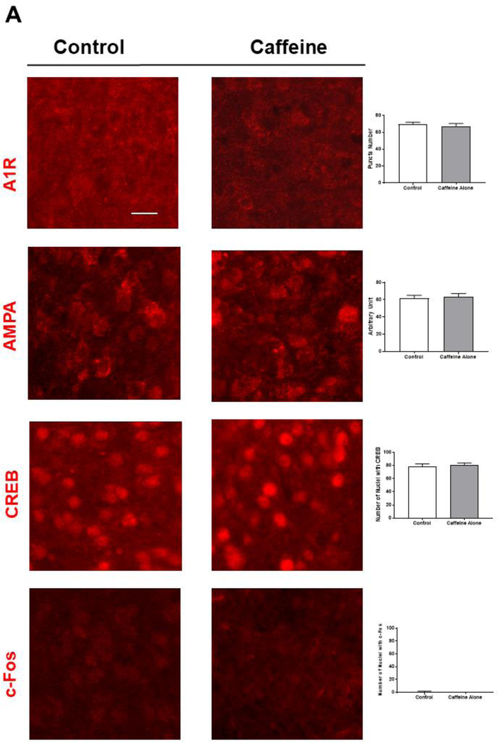
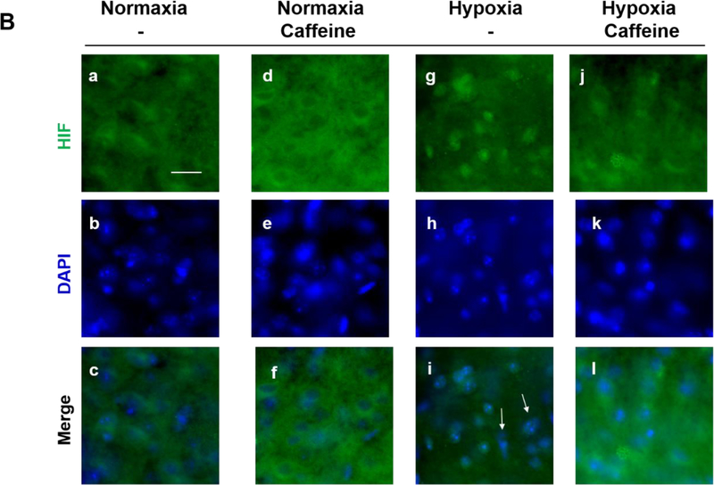
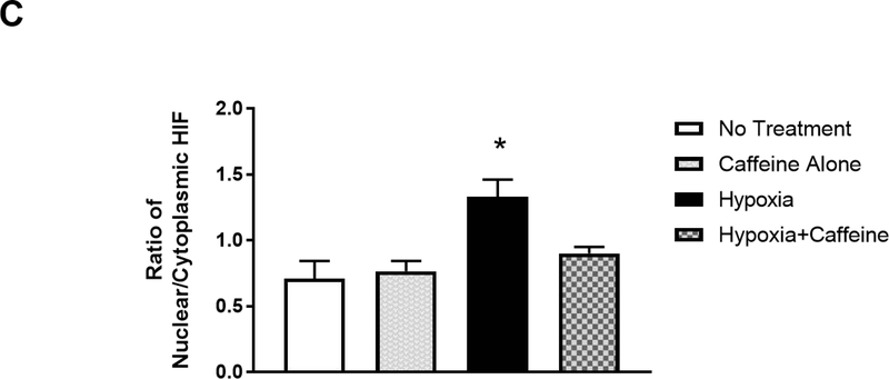
Figure 4.
Caffeine blocked hypoxia-induced nuclear accumulation of HIF1-α in vivo. (A) Immunostaining for A1R, AMPA receptors, CREB-1 and c-Fos showed that caffeine treatment has minimal/no effect on the expression of these factors in neonatal cortex (n=5). Scale bar, 20 μm. The quantitative results in control and caffeine-treated groups are shown on the right; the number of A1R (70±1.15, 67.3±1.86) or AMPA receptor puncta (67.6±2, 63.6±2), the number of nuclear CREB-1 (79±2.1, 81.3±1.5) or c-Fos (1±0.58, 1±0.01). (B) Neonatal mouse pups were gavage fed with caffeine or vehicle from P4-P7. Subsequently, pups were placed under normoxic (room air) or hypoxic (8% O2) conditions for 20 min prior to cardiac perfusion and isolation of their brains. Brain sections were immunostained with anti-HIF-1α (green, top row) and counter-stained with DAPI to show nuclei (blue, middle row). The overlaid images of HIF-1α immunostaining and DAPI staining are shown in the bottom row. Representative cortical images are shown here. Under normoxic conditions, HIF-1α primarily distributed in the cytoplasm of cortical neurons whether mouse pups received caffeine therapy or not (Lane 1, a. b. c. and Lane 2, d, e, f). In pups without caffeine therapy, hypoxia induced nuclear accumulation of HIF-1α (Lane 3, g, h, i). White arrows indicate co-localization of HIF-1α and DAPI. Pre-treatment with caffeine prevented subsequent hypoxia-induced nuclear accumulation of HIF-1α in neonatal cortex (Lane 4, j, k, l). Scale bar, 20 μm. (C) Quantification of the ratio of nuclear vs cytoplasmic HIF-1α in neonatal cortex in each group (control, 0.71±0.05; caffeine alone, 0.77±0.03; hypoxia, 1.33±0.05; hypoxia and caffeine, 0.9±0.02, p<0.001, n=5)


