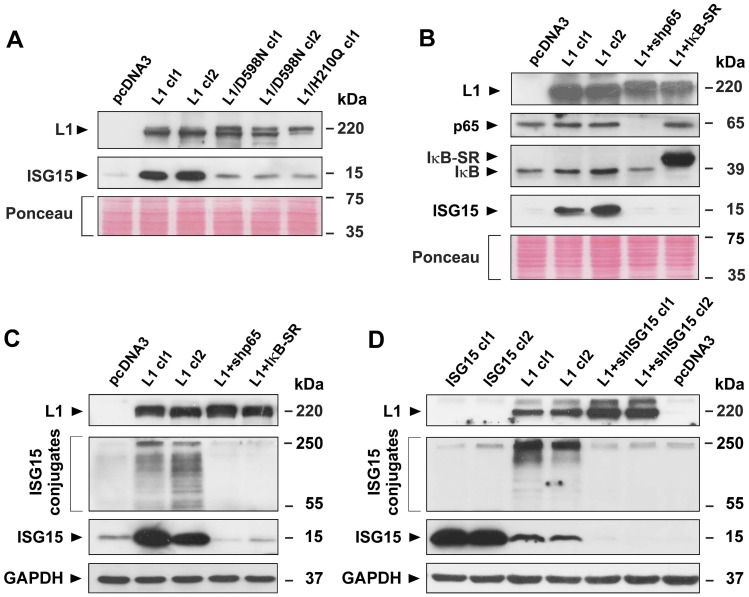
Figure 4. Induction of ISG15 and ISGylation by L1 in CRC cells is blocked when NF-κB signaling is inhibited, or when point mutant L1 forms are expressed in cells.
(A) LS 174T CRC cell clones stably expressing the pcDNA3 control plasmid, or L1 (L1 cl1 and cl2), or the mutant forms of L1 (L1/D598N cl1 and cl2 and L1/H210Q cl1) were analyzed for ISG15 expression by western immunoblotting. (B) CRC cell clones stably expressing the control plasmid pcDNA3, L1 and L1+shRNA to p65 (L1+shp65), or L1 and the IκBα super repressor (IκB-SR) were analyzed for the expression of p65, IκB-SR, IκB, and ISG15 with the relevant antibodies. (C) ISGylation and the levels of free ISG15 were determined in the CRC cell clones described in (B) by western blotting using antibodies to ISG15. (D) ISGylation and free ISG15 levels were determined in CRC cell clones overexpressing ISG15, L1 and L1+shRNA to ISG15. Ponceau staining and GAPDH levels served as markers for equal loading of the gels.

