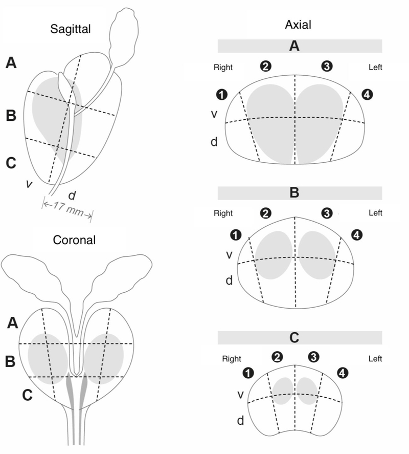
Figure 2.
The 24-sector template, based on the Swedish National PC Guidelines, used to record the anatomical localization of the MRI-lesions and the tumour localization on the whole mount sections. The index tumours were considered detected if there was a lesion with a PI-RADS score 3–5 described in the same or neighbouring sector (approximate match). A = base, B = mid-gland, C = apex v = ventral, d = dorsal

