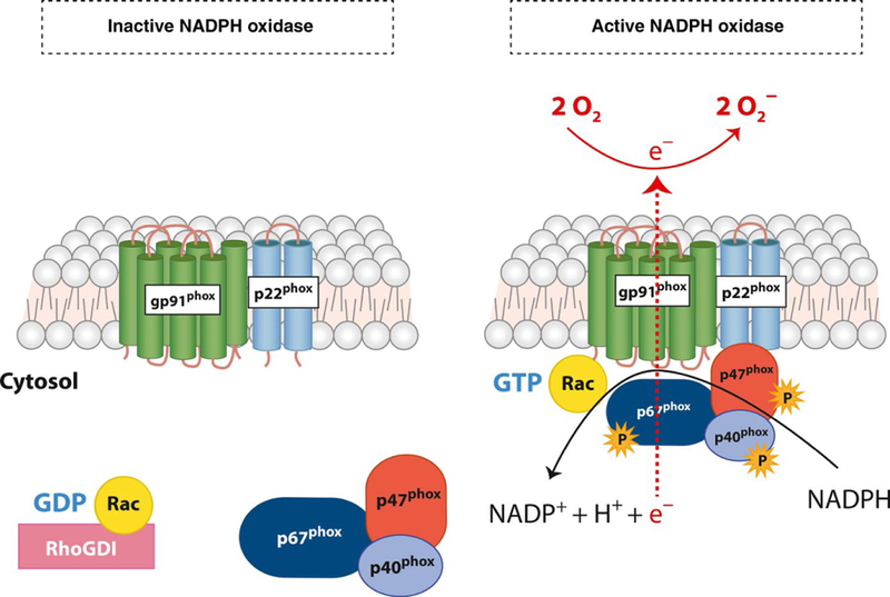
FIGURE 1.
NADPH oxidase structure and assembly. The membrane-restricted heterodimer of NADPH oxidase is comprised of gp91phox and p22phox subunits. This heterodimer is known as flavocytochromeb558. In an inactive state, the cytosolic subunits p67phox, p47phox, and p40phox remain in the cytosol in a self-inhibitory confirmation. On activation, cellular kinases induce the phosphorylation of cytosolic subunits, releasing the inhibitory confirmation. p67phox, p47phox, and p40phox translocate to the membrane along with Rac-GTP and bind to the flavocytochromeb558 forming an active enzyme complex. NADPH-derived electrons are transferred to the substrate molecular oxygen (O2) generating superoxide (O2−) on the other side of the membrane.

