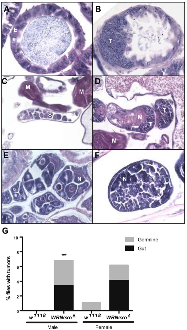
Figure 2: Aged WRNexoΔ mutants have increased tumor incidence.
A) Transverse midgut sections of 35 day-old w1118 controls show epithelial cells (E) with mild to moderate variation in nuclear size and shape, which is a common feature of gut epithelial cells in aging flies. B) In contrast, WRNexoΔ flies show small pleomorphic tumor cells (T) that infiltrate the gut wall and form a mass that protrudes into the lumen. Residual normal gut epithelial cells are present on the right. C) Normal testis in a 35-day old w1118 male sparsely populated with immature spermatocytes and spermatogonia (S) as well as mature spermatozoa (M). D) A 35-day old WRNexoΔ male showing tumor cells (T) in the testes. E) Example of normal follicles from the ovary of a 35 day-old wild-type fly. Normal nurse cells (N) are present within each follicle. F) Section through an abnormal follicle from a 35 day-old WRNexoΔ mutant fly. There is a reduction in nurse cells and the follicle is filled with small, pleomorphic tumor cells (T) whose morphology is reminiscent of germline stem cells. G) Higher total tumor incidence was observed in 35 day-old WRNexoΔ males (p = 0.0029 (males) and 0.067 (females) by Fisher’s exact test. Male: w1118 n = 123, WRNexoΔ n = 118; Female: w1118 n = 94, WRNexoΔ n = 195.

