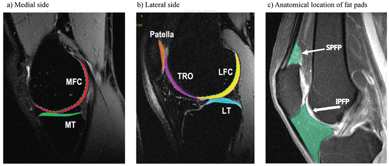
Figure 1:

depiction of the 6 cartilage compartments used to quantify MR-based T1ρ and T2 cartilage relaxation time measurements (a and b) as well as of knee fat pads (c).
a: sagittal 3 T MR image of the medial aspect of the knee showing the cartilage of the medial femoral condyle (MFC) and of the medial tibia (MT).
b: sagittal 3 T MR image of the lateral aspect of the knee showing the cartilage of the lateral femoral condyle (LFC), the lateral tibia (LT), the trochlea (TRO) and the patella (PAT).
c: Representative sagittal MR image of the knee showing the anatomical location of the suprapatellar fat pad (SPFP) and of the infrapatellar Hoffa fat pad (IPFP). Both fat pads are highlighted in green. The arrow with the larger tip points to the suprapatellar fat pad (SPFP). The arrow with the smaller tip points towards the infrapatellar fat pad (IPFP).
MR=magnetic resonance.
