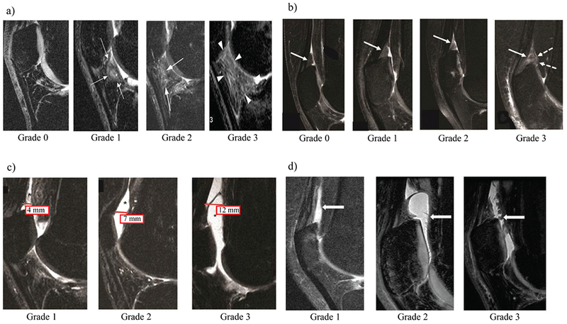
Figure 2.

Illustration and description of the MR-based fat pad-synovitis grading scheme used in this study. Four subfeatures were scored on every knee MR scan. (a) The degree of infrapatellar Hoffa fat pad abnormality (IPFP abnormality) was scored on mid-line sagittal FS CUBE and non-fat saturated PD images according to the Anterior Cruciate Ligament OsteoArthritis Score (ACLOAS)31. White arrows point towards abnormal findings of the IPFP at each grade. Grade 0; normal appearing fat pad, only small physiologic vascular structures visible Grade 1; mild hyperintensity of the fat pad, Grade 2; moderate fat pad hyperintensity, Grade 3; severe hyperintense signal changes within the fat pad. (b) The degree of suprapatellar fat pad abnormality (SPFP abnormality) was assessed on sagittal fat-saturated intermediate weighted sequences with a small modification to a previously published grading system32. White arrows show SPFP. Grade 0 was defined by a normal appearing SPFP with isointense signal compared to the prefemoral fat pad. Grade 1 was defined by mild, hyperintense signal alterations in the SPFP compared to the prefemoral fat pad. Grade 2 was defined by moderate hyperintense signal alterations within the SPFP relative to the prefemoral fat pad signal intensity. Grade 3 SPFP was defined by severely hyperintense signal alterations within the SPFP which were accompanied by extensive fraying and/or mass effect of the fat pad. (c) The extent of effusion-synovitis was scored by assessing the anterior-posterior diameter of the joint effusion in mm on sagittal MR-images as described before. In detail, this standardized scoring system was applied to sagittal fat saturated CUBE images in the lateral compartment just mesial to the fibular head unless there was evidence of patellar subluxation: in this case, a mid-fibular head section was used. The suprapatellar recess was used as the point of reference. In detail, effusion-synovitis was graded from 0 to 3 according to the degree of capsular distension with grade 0 being equivalent to a <2 mm anterior-posterior diameter of the effusion. A joint effusion spanning ≥2 and <5 mm in the anterior-posterior (ap) diameter on the mid-slice sagittal image was graded as 1, while a joint effusion between ≥5 and <10 mm was graded as 2. Any effusion measuring equal or more than 10 mm in the ap-diameter was scored as grade 3. (d) Synovial proliferation grading scheme. White arrows point towards synovial proliferations. The presence and severity of synovial proliferations was assessed on sagittal fat saturated CUBE and non-fat saturated PD images in the suprapatellar recess and other visible areas of the joint. Grade 1 corresponded to a smooth synovium, with no proliferation or synovial bands visible; grade 2 was defined as a mild irregularity of the synovium, either focal or diffuse, and the presence of some synovial bands or small bodies; grade 3 was defined as extensive synovial thickening with irregular villo-nodular proliferation. MR = Magnetic Resonance; ACLOAS = Anterior Cruciate Ligament OsteoArthritis Scoring as published by Roemer et al31.
