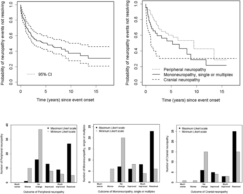
Figure 2:
Physician determined change in peripheral neuropathy, mononeuropathy or cranial neuropathy (n=149) attributed to SLE and non-SLE using attribution model B. Top panel: Survival curves for resolution of all neuropathies (left) and individual neuropathies (right). Lower panel: Likert scale scores for physician assessment of outcome over the duration of followup are shifted to the right indicated improvement and this is most pronounced for cranial neuropathies (right).

