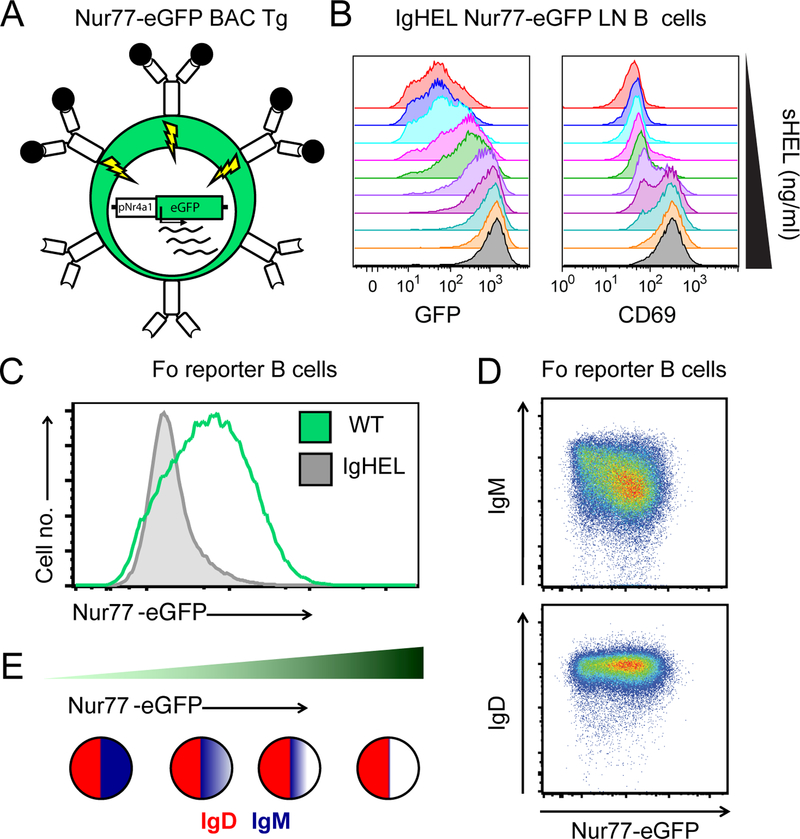
Figure 1. Nur77-eGFP BAC Tg reporter of antigen receptor signaling marks self-reactive B cells in vivo.
A. Schematic of Nur77-eGFP BAC Tg depicts eGFP transcript under the control of the regulatory region of Nr4a1. Since Nr4a1 is a primary response gene (PRG) that is rapidly transcribed in response to antigen receptor signaling, antigen encounter results in rapid GFP induction in reporter B cells.
B. IgHEL BCR Tg B cells harboring the Nur77-eGFP BAC Tg were stimulated in vitro with varying doses of soluble HEL antigen, and both GFP and CD69 expression were assessed via FACS after 24 hours.
C. Mature follicular (Fo) splenic B cells (CD23HICD93-B220+) from Nur77-eGFP BAC Tg mice with or without IgHEL BCR Tg (in the absence of cognate HEL Ag) were assessed for GFP expression via FACS immediately ex vivo. Restricting the BCR repertoire to restrict endogenous antigen recognition markedly reduces GFP expression.
D. Surface IgM and IgD BCR expression on mature Fo splenic B cells from Nur77-eGFP BAC Tg mice was assessed via FACS, revealing that IgM expression but not IgD expression is inversely correlated with GFP. Dynamic range of IgM across the repertoire is large, varying by more than an order of magnitude along a log10 scale.
E. Schematic depicts relative surface expression of IgM (blue) and IgD (red) BCRs on B cells with increasing expression of GFP (and therefore increasing self-reactivity) across the mature Fo B cell repertoire.

