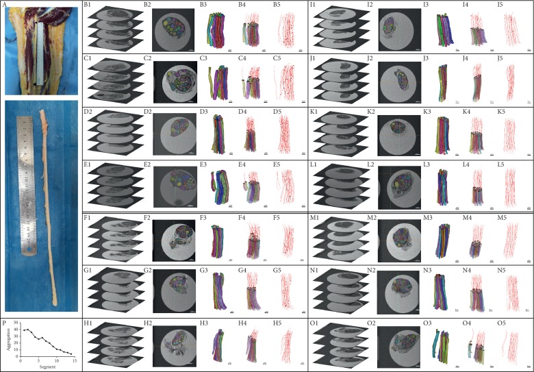
Figure 2.
Analysis of morphological cross fusion of long tibial nerve fascicles. A: tibial nerve full-length specimen intercepted. B to O images showed 14 two-centimetre nerve samples taken from proximal end to distal end. In each box from left to right (1–5): Micro-MRI scan images are displayed in each group. In the two-dimensional image, the nerve fascicles were segmented, the nerve fascicles were reconstructed in three dimensions, and the central line was fitted to the three-dimensional reconstruction model. P: the change of the tibial nerve from sciatic nerve branches to the popliteal fossa: cross fusion of the nerve fascicles decreased gradually from proximal to distal. B2 to O2: scale bar 1 mm; B3 to O3, B4 to O4, and B5 to O5: scale bar 2 mm.

