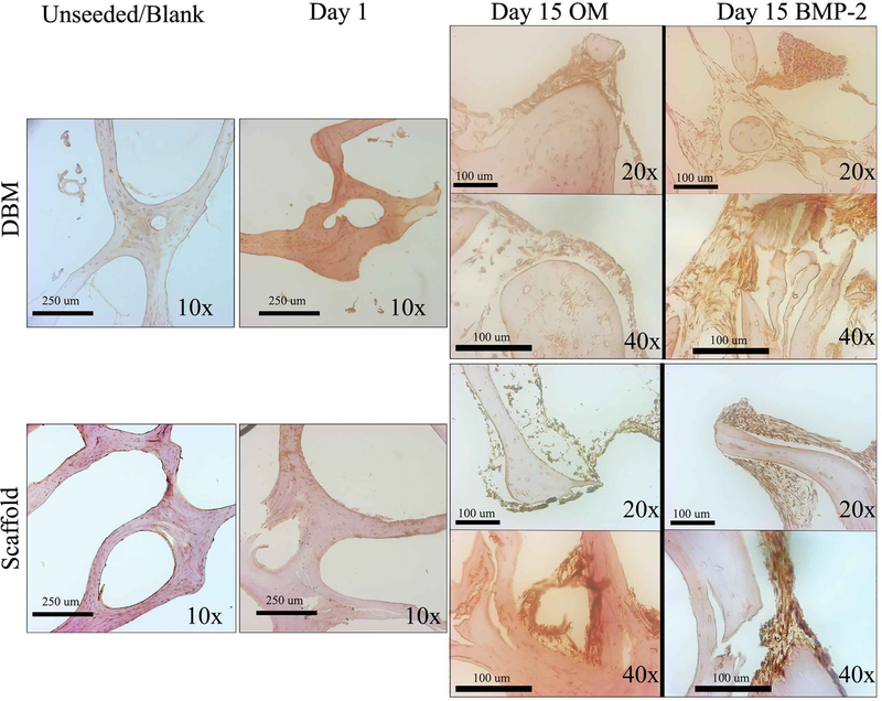
Figure 6. ALP expression increases over time in both DBM and scaffolds.
DBM (top panels) or decellularized bone (bottom panels) scaffolds were sectioned unseeded, after 1-day culture of C2C12 cells or after 15 days in OM or OM with 100 ng/mL BMP-2. Sections were stained for ALP expression by immunohistochemistry, and representative images are shown at 10X, 20X, and 40X. Images were taken at different locations and did not represent subsets of each other.

