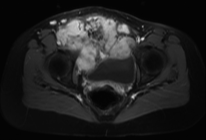
Figure 1.

Representative axial image of a gadolinium‐enhanced MRI of the pelvis in a 21‐y‐old woman with desmoid fibromatosis of the right hemipelvis. The tumor displaced the bladder and uterus and involved the superior pubic ramus, lymphatics, and common femoral and external iliac veins
